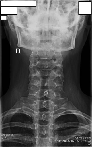
AP CERVICAL
Anteroposterior cervical spine projection protocol
Exposure Factors
Medium exposure: Parameters for optimal visualization of cervical vertebrae
Anatomical Structures Visible
Should be clearly observed:
- Vertebral bodies from C3 to C7
- Spinous processes
- Uncinate processes
- Transverse processes
- Intervertebral disc spaces
- Pedicles
- Skull base
- First thoracic vertebra (T1)
Cassette Size and Orientation
Longitudinal orientation to cover entire cervical spine
Patient Positioning
Central Ray Point
Location: Directed to the fourth cervical vertebra (C4)
Angulation: 15 to 20° cephalad direction
Patient with limitation: If unable to raise chin, increase angulation to 30-40°
Optimal Image Characteristics
Vertebrae C3-C7
Clearly visible
Disc Spaces
Intervertebral spaces open
Symmetry
Bilateral structures aligned
Skull Base and T1
Included in field
Common Technical Challenges
Frequent problems in AP cervical projection:
- Mandible-vertebrae superimposition from insufficient chin elevation
- Shoulders not lowered obscuring lower cervical vertebrae
- Head rotation causing spinous process asymmetry
- Poor angulation failing to open intervertebral spaces
Solution: Verify occlusal plane perpendicular to cassette and shoulders completely lowered
Patient Instructions
"Hold your breath during the exposure"
Maintain position without movement during radiographic exposure
CERVICAL TRAUMA CONSIDERATIONS
In patients with suspected cervical trauma or immobilized:
- Perform in supine position without moving patient
- Place cassette directly behind patient
- If cervical collar present, do not remove except by medical indication
- Angulation according to patient's mobilization capability
Priority: Patient safety over image quality in trauma cases
Technical Variations
Obese Patient
Increase kV and mAs according to thickness adjustment chart.
Geriatric Patient
Possible kyphosis requires greater cephalic angulation.
Pediatric Patient
Reduce exposure according to age and ALARA protocol.